 | Info
Sheets |
| | | | | | | | | | | | | | | | | | | | | | | | |
 | Out-
side |
| | | | |
|
| | | | |
Result : Searchterm 'Voxel Volume' found in 0 term [ ] and 1 definition [ ] and 1 definition [ ], (+ 15 Boolean[ ], (+ 15 Boolean[ ] results ] results
| 1 - 5 (of 16) nextResult Pages :  [1] [1]  [2 3 4] [2 3 4] |  | |  | Searchterm 'Voxel Volume' was also found in the following service: | | | | |
|  |  |
| |
|
( SNR or S/N) The signal to noise ratio is used in MRI to describe the relative contributions to a detected signal of the true signal and random superimposed signals ('background noise') - a criterion for image quality.
One common method to increase the SNR is to average several measurements of the signal, on the expectation that random contributions will tend to cancel out. The SNR can also be improved by sampling larger volumes (increasing the field of view and slice thickness with a corresponding loss of spatial resolution) or, within limits, by increasing the strength of the magnetic field used. Surface coils can also be used to improve local signal intensity. The SNR will depend, in part, on the electrical properties of the sample or patient being studied.
The SNR increases in proportion to voxel volume (1/resolution), the square root of the number of acquisitions ( NEX), and the square root of the number of scans ( phase encodings). SNR decreases with the field of view squared (FOV2) and wider bandwidths. See also Signal Intensity and Spin Density.
Measuring SNR:
Record the mean value of a small ROI placed in the most homogeneous area of tissue with high signal intensity (e.g. white matter in thalamus). Calculate the standard deviation for the largest possible ROI placed outside the object in the image background (avoid ghosting/aliasing or eye movement artifact regions).
The SNR is then:
Mean Signal/Standard Deviation of Background Noise | | | |  | | | | • Share the entry 'Signal to Noise Ratio':    | | | | | | | | | |  Further Reading: Further Reading: | | Basics:
|
|
News & More:
| |
| |
|  | |  |  |  |
| |
|
Device Information and Specification
CLINICAL APPLICATION
Whole body
CONFIGURATION
Cylindrical Wide Short Bore
Opt. (WIP) Single and Multi Voxel
SE, FE, IR, FastSE, FastIR, FastFLAIR, Fast STIR, FastFE, FASE, Hybrid EPI, Multi Shot EPI; Angiography: 2D(gate/non-gate)/3D TOF, SORS-STC
IMAGING MODES
Single, multislice, volume study
TE
8 msec min. SE; 1.2 msec min. FE
less than 0.015 (256x256)
1.0 min. 2-DFT: 0.2 min. 3-DFT
32-1024, phase;; 64-1024, freq.
65.5 cm, patient aperture
4050 kg (bare magnet incl. L-He)
COOLING SYSTEM TYPE
Closed-loop water-cooled
Liquid helium: approx. less than 0.05 L/hr
Passive, active, auto-active
| |  | |
• View the DATABASE results for 'Excelart AG™ with Pianissimo' (2).
| | | | |
|  | |  |  |  |
| |
|

(Signa VH/i 3.0T)
With GE Healthcare
leading-edge technology in ultra-high-field imaging. The 3 T VH/i provides a platform for advanced applications in radiology, cardiology, psychology and psychiatry. Real-time image processing lets you acquire multislice whole brain images and map brain functions for research or surgical planning. And the 3 T Signa VH/i is flexible enough to provide clinicians with high performance they require. It can provide not only outstanding features in brain scanning and neuro-system research, but also a wide range of use in scanning breasts, extremities, the spine and the cardiovascular systems.
Device Information and Specification CLINICAL APPLICATION Whole body
T/R quadrature head, T/R quadrature body, T/R phased array extremity (opt) SE, IR, 2D/3D GRE, FGRE, RF-spoiled GRE, FSE, Angiography: 2D/3D TOF, 2D/3D phase contrast vascular IMAGING MODES Single, multislice, volume study, fast scan, multi slab, cine, localizer 100 Images/sec with Reflex100 MULTISLICE 100 Images/sec with Reflex100 2D 0.5-100mm in 0.1mm incremental 128x512 steps 32 phase encode H*W*D 260cm x 238cm x 265cm POWER REQUIREMENTS 480 or 380/415, 3 phase ||
COOLING SYSTEM TYPE Closed-loop water-cooled grad. Less than 0.14 L/hr liquid He | |  | |
• View the DATABASE results for 'Signa 3.0T™' (2).
| | | | |
|  |  | Searchterm 'Voxel Volume' was also found in the following service: | | | | |
|  |  |
| |
|

From GE Healthcare;
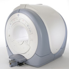
The Signa HDx MRI system is GE's leading edge whole body magnetic resonance scanner designed to support high resolution, high signal to noise ratio, and short scan times.
Signa HDx 3.0T offers new technologies like ultra-fast image reconstruction through the new XVRE recon engine, advancements in parallel imaging algorithms and the broadest range of premium applications. The HD applications, PROPELLER (high-quality brain imaging extremely resistant to motion artifacts), TRICKS (contrast-enhanced angiographic vascular lower leg imaging), VIBRANT (for breast MRI), LAVA (high resolution liver imaging with shorter breath holds and better organ coverage) and MR Echo (high-definition cardiac images in real time) offer unique capabilities.
Device Information and Specification CLINICAL APPLICATION Whole body
CONFIGURATION Compact short bore SE, IR, 2D/3D GRE, RF-spoiled GRE, 2DFGRE, 2DFSPGR, 3DFGRE, 3DFSPGR, 3DTOFGRE, 3DFSPGR, 2DFSE, 2DFSE-XL, 2DFSE-IR, T1-FLAIR, SSFSE, EPI, DW-EPI, BRAVO, Angiography: 2D/3D TOF, 2D/3D phase contrast vascular IMAGING MODES Single, multislice, volume study, fast scan, multi slab, cine, localizer H*W*D 240 x 2216,6 x 201,6 cm POWER REQUIREMENTS 480 or 380/415, 3 phase ||
COOLING SYSTEM TYPE Closed-loop water-cooled grad. | |  | | | |
|  | |  |  |  |
| |
|
(CSI) Chemical shift imaging is an extension of MR spectroscopy, allowing metabolite information to be measured in an extended region and to add the chemical analysis of body tissues to the potential clinical utility of Magnetic Resonance. The spatial location is phase encoded and a spectrum is recorded at each phase encoding step to allow the spectra acquisition in a number of volumes covering the whole sample. CSI provides mapping of chemical shifts, analog to individual spectral lines or groups of lines.
Spatial resolution can be in one, two or three dimensions, but with long acquisition times od full 3D CSI. Commonly a slice-selected 2D acquisition is used. The chemical composition of each voxel is represented by spectra, or as an image in which the signal intensity depends on the concentration of an individual metabolite. Alternatively frequency-selective pulses excite only a single spectral component.
There are several methods of performing chemical shift imaging, e.g. the inversion recovery method, chemical shift selective imaging sequence, chemical shift insensitive slice selective RF pulse, the saturation method, spatial and chemical shift encoded excitation and quantitative chemical shift imaging.
See also Magnetic Resonance Spectroscopy. | |  | |
• View the DATABASE results for 'Chemical Shift Imaging' (6).
| | | | |  Further Reading: Further Reading: | Basics:
|
|
News & More:
| |
| |
|  | |  |  |
|  | 1 - 5 (of 16) nextResult Pages :  [1] [1]  [2 3 4] [2 3 4] |
| |
|
| |
 | Look
Ups |
| |